Introduction
A 76-year-old woman presented with increasing hoarseness, odynophagia, and otalgia for the last 6 months. She had been seen by a number of different physicians. The results of bronchoscopic examination and barium swallow were unrevealing. A chest CT performed at another institution showed that the left hemidiaphragm was elevated, and that the left lung was smaller and more lucent than the right lung.
She successfully quit smoking 3 years earlier. She had undergone left lung upper lobectomy 20 years ago for hemoptysis but no diagnosis was given. Other significant medical history included palpitations, angina, and carotid endarterectomy. Findings on physical examination were noncontributory.
She was referred for FDG-PET scanning to further assess her condition. For ease of image interpretation, the relevant images of the subsequently obtained separate contrast-enhanced CT of the chest and the neck (all performed within 2 weeks after the FDG-PET) are shown first, followed by the appropriate FDG PET images.

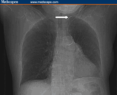
Scout image of a chest CT scan (14 days after FDG-PET) shows elevated left hemidiaphragm and clips in the left lung apex (white arrow) consistent with prior left upper lobectomy.

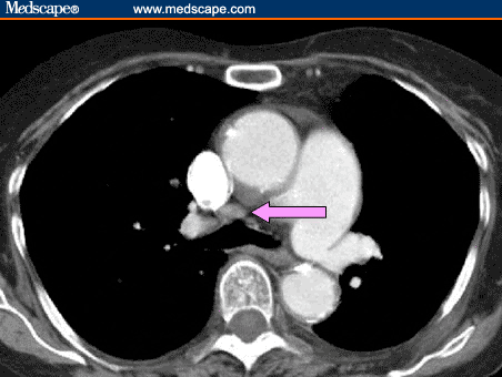
Axial contrast-enhanced chest CT shows mild precarinal lymphadenopathy (purple arrow).

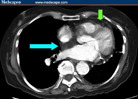
Axial contrast-enhanced chest CT at a lower level shows thickened interatrial septum of largely fat attenuation (blue arrow) and rather prominent epicardial fat (green arrow).

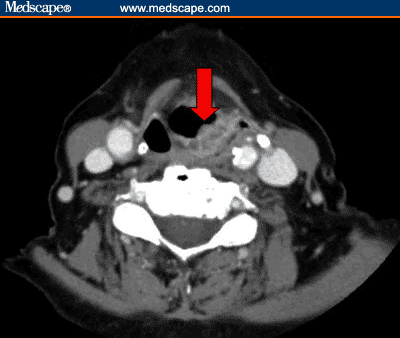
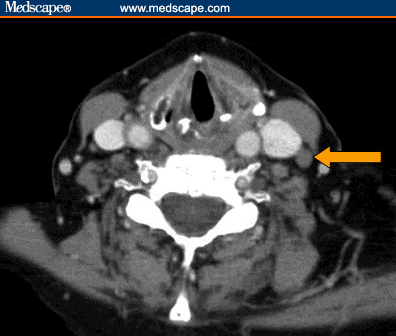
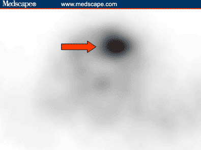
Axial neck CT (10 days after FDG-PET) shows a contrast-enhancing supraglottic mass (red arrow) extending from almost the whole of the left aryepiglottic fold down to the superior aspect of the left false vocal cord. There is likely involvement of the left posterolateral pharyngeal wall.

Axial contrast-enhanced neck CT (10 days after FDG-PET) shows a borderline enlarged level III lymph node (orange arrow) at the level of the lower border of the left supraglottic mass. There are no other enlarged cervical lymph nodes outside this region.

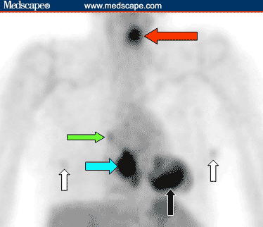
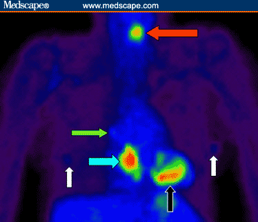
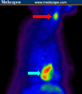
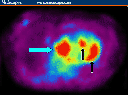
Anterior projection image of whole-body FDG-PET shows a large area of intense FDG uptake in the lower mediastinum (blue arrow). There is also a focus of intense activity in the left laryngeal region (red arrow). There is focal low-grade uptake at the right superior cardiac border, considered to represent physiologic uptake in the right atrium (green arrow). Physiologic uptake is noted in the ventricular myocardium (black arrow) and in the breasts (white arrows).

Anterior projection image of whole-body FDG-PET shows a large area of intense FDG uptake in the lower mediastinum (blue arrow). There is also a focus of intense activity in the left laryngeal region (red arrow). There is focal low-grade uptake at the right superior cardiac border, considered to represent physiologic uptake in the right atrium (green arrow). Physiologic uptake is noted in the ventricular myocardium (black arrow) and in the breasts (white arrows).

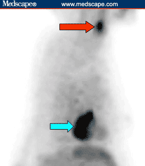
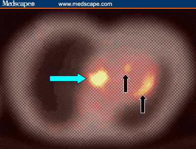
Lateral projection image of whole-body FDG-PET shows the focal increased FDG activity in the lower mediastinum (blue arrow) and in the neck (red arrow).

Lateral projection image of whole-body FDG-PET shows the focal increased FDG activity in the lower mediastinum (blue arrow) and in the neck (red arrow).
Figure 8. Cine format of FDG-PET projection images.
Figure 8. Cine format of FDG-PET projection images.

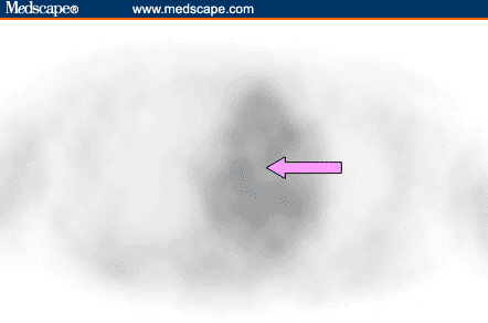
Axial FDG-PET image (at approximately the same level as the axial chest CT in Figure 2) shows no abnormal FDG uptake in the precarinal region (purple arrow).

Axial FDG-PET image (at approximately the same level as the axial chest CT in Figure 2) shows no abnormal FDG uptake in the precarinal region (purple arrow).

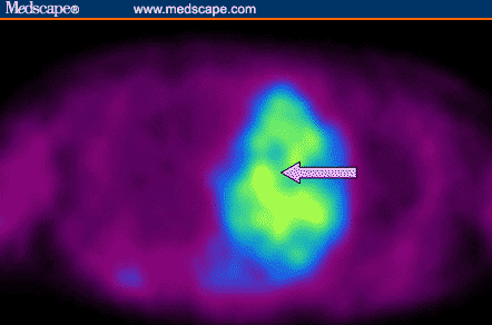
Axial FDG-PET image (at approximately the same level as the axial chest CT in Figure 3) shows intense FDG uptake corresponding to the thickened interatrial septum (blue arrow). Physiologic left ventricular myocardial FDG uptake is noted (black arrows).

Axial FDG-PET image (at approximately the same level as the axial chest CT in Figure 3) shows intense FDG uptake corresponding to the thickened interatrial septum (blue arrow). Physiologic left ventricular myocardial FDG uptake is noted (black arrows).

Fusion image between the axial transmission image (used for attenuation correction of PET images) and the axial emission FDG-PET image in Figure 10 confirms the interatrial septal location of the intense FDG uptake (blue arrow). Physiologic left ventricular myocardial FDG uptake is noted (black arrows). The transmission and emission images were acquired at the same level and the same bed position.

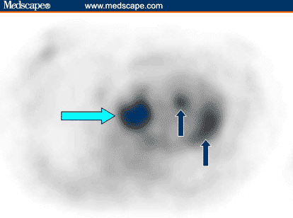
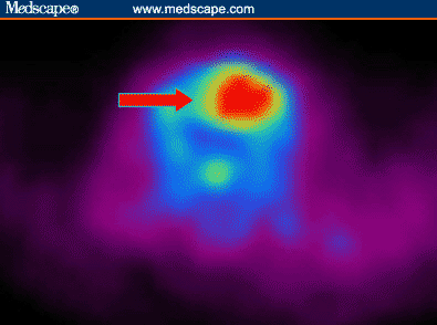
Axial FDG-PET image (at approximately the same level as the axial neck CT in Figure 4) shows focal intense FDG uptake in the lateral aspect of the left laryngeal region corresponding to the supraglottic mass at this site (red arrow). There are no other FDG-avid foci in the neck, including in the region of the borderline level III lymph node on the left (as seen in Figure 5).

Axial FDG-PET image (at approximately the same level as the axial neck CT in Figure 4) shows focal intense FDG uptake in the lateral aspect of the left laryngeal region corresponding to the supraglottic mass at this site (red arrow). There are no other FDG-avid foci in the neck, including in the region of the borderline level III lymph node on the left (as seen in Figure 5).
Question
In this case, the clinical history and imaging findings were compatible with a diagnosis of:
Positron emission tomography using F-18 fluoro-deoxyglucose (FDG-PET) assists in the differentiation of many malignant tissues that have elevated glycolysis from normal tissues. FDG uptake is not tumor-specific, as benign processes such as inflammatory lesions, active muscular activity, or brown adipose tissue (BAT) can exhibit increased FDG activity. Briefly, BAT is abundantly present in infancy and diminishes with age.[1] BAT expresses the unique mitochondrial uncoupling protein (UCP) that permits direct heat generation in response to cold exposure (nonshivering thermogenesis), ingestion of food (diet-induced thermogenesis),[2] or increased sympathetic activity.[3] This process requires increased glycolytic metabolism to provide adenosine triphosphate (ATP) that is necessary for fatty acid oxidation.[1] With routine use of PET/CT fusion imaging, it is now well recognized that increased FDG activity can localize to normal adipose tissue (most likely BAT) typically in the neck, supraclavicular regions, axillae, and thoracic paravertebral intercostal space.[1] Increased FDG uptake attributable to BAT has been described in the mediastinum (among the large vessels) and in the perinephric fat.[1] This phenomenon appears to occur more often in females,[1] younger persons,[1] during the colder months,[4] possibly in those with lower body mass index,[3] and possibly in those with anxiety.[1] Intense FDG uptake has also been described in hibernoma (encapsulated benign tumor of BAT).[5]
The exact etiology of the FDG-avid tissue in the interatrial septum is uncertain because histopathologic sampling was not performed. On the basis of the available imaging features and clinical data, the authors believe that the most plausible explanation for this unusual finding is lipomatous hypertrophy of the interatrial septum (LHIS) with high BAT content.
LHIS, also termed "massive fatty deposits" or "lipomatous hamartoma", is an uncommon disorder of unknown etiology characterized by fat accumulation in the interatrial septum.[6] The diagnosis is usually made incidentally. It typically occurs in elderly, obese patients and may cause arrhythmia.[6] LHIS generally has thickness > 2 cm (normal thickness < 1 cm)[7] and can be associated with increased epicardial and/or mediastinal fat.[6] Multislice CT is a useful method to diagnose LHIS, typically showing a mass of fat attenuation with sharp margins. A dumbbell-shaped configuration is a characteristic finding (due to sparing of the fossa ovalis) on 3-D reconstruction, although this feature may not be present in all axial CT images.[6] Histologically, LHIS typically shows unencapsulated hyperplasia of mature multivacuolated fat cells with variable amounts of BAT, entrapped myocytes, and fibrosis.[6] High BAT in LHIS has been reported in one histopathologic study.[8]
The differentiation of LHIS from other cardiac neoplasms can be difficult based on noninvasive conventional imaging alone. In the current clinical context, other diagnostic possibilities are much less likely. Briefly, myxomas are the most common primary cardiac tumors. The majority are solitary, intracavitary, heterogenous, hypoattenuated masses in the atria on CT.[6,9] Most myxomas are pedunculated on a fibrovascular stalk, thus differentiating it from LHIS.[6] Cardiac lipomas are encapsulated and occur in a slightly younger age group (< 57 years of age).[6] To our knowledge, interatrial hibernoma (an encapsulated structure) has not been described. Rhabdomyomas and fibromas are common benign cardiac tumors in infants and children and usually occur in the ventricles.[6] Cardiac liposarcoma is a rare, rapidly growing tumor that predominantly appears in the right atrium and usually with early signs of local invasion and hemodynamic compromise.[6] Metastatic malignancy is extremely unlikely in this case given the lack of abnormal soft tissue density at the interatrial septum on CT and the relatively long disease-free period (> 2 years) following chemoradiation therapy directed mainly to treat locally advanced head and neck cancer.
The vast majority of head and neck malignancies are squamous cell carcinoma of the nasopharynx, oropharynx, oral cavity, and larynx (HNSCC). Most data on the utility of FDG-PET in head and neck cancer apply to HNSCC. Early-stage HNSCC is usually treated by surgery or radiation therapy as a single treatment with generally good results and similar cure rates.[10] Advanced-stage HNSCC usually requires multimodality therapy such as surgery, chemotherapy, and/or radiation therapy. FDG-PET may be of value in the localization of a primary tumor in the uncommon setting of metastatic squamous cell carcinoma in a cervical lymph node of unknown primary.[11] Once the primary tumor has been identified, FDG-PET alone has little role in the T-staging due to the lack of anatomic details such as the extent of local invasion or the proximity to adjacent major structures. FDG-PET is equivalent or superior to conventional anatomic imaging modalities in the detection of nodal metastases. FDG-PET is particularly valuable in the detection of metastatic disease in normal-sized lymph nodes as small as 0.6 cm.[12] Distant metastases from HNSCC are uncommon at diagnosis (< 5%).[11] Preliminary data suggest a potential for FDG-PET in the detection of occult distant metastases, particularly in the mediastinal lymphatics in patients with advanced-stage HNSCC.[13] Whole-body FDG-PET may also be important in the diagnosis of synchronous primary tumors.[11]
FDG-PET is best interpreted with anatomic correlation to minimize false-positive results from increased FDG uptake in benign processes, such as in the interatrial septum in this case. The exact status of the small, nonpalpable left level III cervical lymph node is unknown. Metastatic disease at this site and in the precarinal region is, however, considered unlikely in view of the absence of FDG avidity and long-term favorable course.
Medscape Radiology. 2005;6(1) © 2005 Medscape
Cite this: PET Case Cavalcade, Case XXVII: Hoarseness in an Older Woman - Medscape - Mar 31, 2005.
Comments